5.3 Radio Immuno Assay (RIA)
In the RIA, IgG subclasses are quantified as immune complexes after binding of radioactively labelled specific antibody. In case of a 'direct' technique, a radioactively labelled anti-IgG subclass-specific antibody is used. In an 'indirect' technique, an anti-IgG subclass-specific antibody directed against the first antibody. Since working with radioactively labelled reagents requires special precautions and is relatively costly, radio immunoassays have been largely replaced by enzyme-linked immunoassays.
5.4 Enzyme-linked Immunosorbent Assay (ELISA)
In this technique, enzyme-labelled antibodies are used for detection. In the 'sandwich'-type of enzyme immunoassay, the IgG subclass of interest is captured by anti-human IgG subclass-specific antibody and quantified by an enzyme-labelled anti-IgG antibody. Because of its high sensitivity and specificity, this assay allows accurate measurement of very low levels of IgG subclasses. In figure 9, a schematic outline of the ELISA technique is shown. The sensitive ELISA comprises many incubation-and washing steps. Because of the need for high dilutions when measuring IgG subclasses in sera, ELISA assays may be less reproducible in comparison with RID and nephelometry. For measurement of IgG and its subclasses in large numbers of samples, ELISA is increasingly being replaced by nephelometry. ELISA may be advocated for measuring IgG subclasses in other body fluids than serum/plasma, e.g. saliva, cerebrospinal fluid and broncho-alveolar lavage fluid.
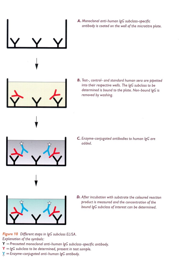
Brief outline of the method (figure 10):
An ELISA is generally performed in wells of microplates.
- Wells of the plates are coated with unlabelled monoclonal antihuman IgG subclass-specific
antibody and washed (figure 10A);
- Test samples, standard-and control sera are introduced in the respective wells
and incubated; The IgG subclass to be determined will bind to the solid phase
and non-bound IgG is removed by washing (figure 10B);
- Enzyme-labelled anti-human IgG antibodies are added to each well and non-bound
conjugate is removed by washing (figure 10C);
- Plates are incubated with substrate solution;
- After incubation, the coloured reaction product is measured photometrically
(figure 10D);
- The concentration of the IgG subclasses in the test samples is calculated
relative to the values of the calibration curve.