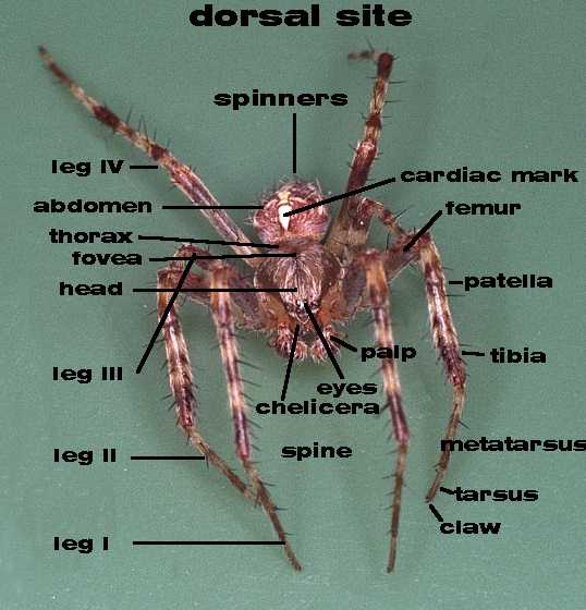
The body of a spider consists of two easily recognizable
parts.
A prosoma or cephalothorax (composed of the fused head and
thorax) and the abdomen (the rear body).
A narrow waist (pedicel) connects both parts.
The upper side of the spider is called the dorsal side (i.e. the
back) and the bottom the ventral side (i.e. the belly).
The cephalothorax at the dorsal side is made of chitin what is called the
carapax.
The back the prosoma is called the abdomen or opisthosoma.
Directly behind the head is a groove, the fovea. The zone just behind
the eyes is called the clypeus (forehead). The part on the dorsal
site between the legs is called sternum. The palps in the
front are used for sensory purposes. In the male the palps are modified for
putting sperm into the epigyne of the female, that is situated on the
underside of the abdomen. The jaws are located below the eyes and are called
chelicera. The piercing parts of these jaws are the fangs that
deliver the venom via a small hole at its tip. The
silk is produced by the silk glands and released by the spinners.
Immediately in front of the spinnerets, in certain spiders, is the
cribellum, a special plate, containing a row of openings through which
many strands of very fine silk is produced. With their hind legs, they can
comb the silk to produce fluffy wool like silk to make the so-called hackle
bands on their webs.
See The spinnerets and the properties of silk
More detailed pages can be found here: Spider information
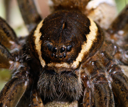
The eight eyes of Dolomedes fimbriatus
The eyes are located in the front. Most spiders have eight eyes, some
families six.
The eyes are singular and are called ocelli, unlike the insects that
have compound eyes. The main pair of eyes is always the middle pair and has
a different construction than the other eyes.
The light sensitive cells point towards the incoming light and is therefore
referred to as indirect retina. Except the crab spiders and jumping spiders,
the main eyes are small or even absent in spiders with six eyes. The
construction is similar to as in humans and these eyes have a high
resolution. In the other eyes the light sensitive cells points away from the
light and have an indirect retina. These secondary eyes give the spider a
much broader field of vision than the main eyes and are used to give them
the ability to judge distance. During hunting, these secondary eyes catch
the movement of the prey and the main eyes are used to focus as it moves
closer to the prey.
The trachea holes and the book lungs are the inlets for oxygen.
These holes are situated at the underside of the abdomen. The number of
trachea and book lungs and their position varies from family to family.
At the front dorsal side of the abdomen is the heart spot located.
Below this spot is the spiders heart. Big spiders have a heart beat around
30 - 70 beats a minute whereas in smaller spiders the heart beat can rise
up to 200 beats per minute.
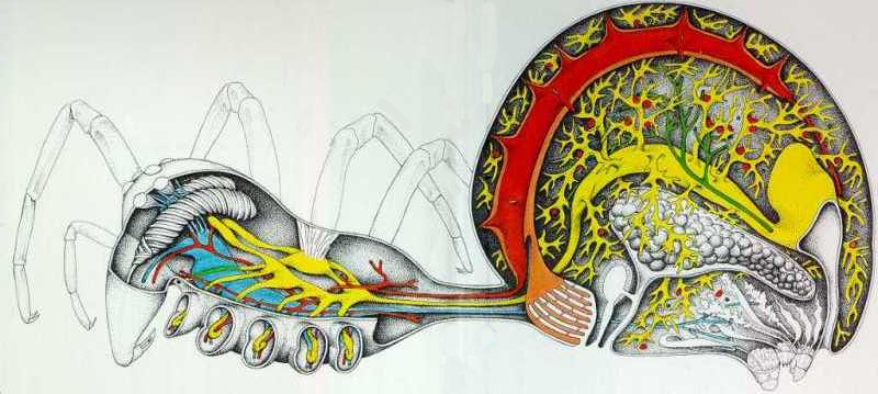
The internal anatomy of a female (picture from reference 7).
Spiders have an alimentary canal (yellow),
a blood vascular system (red),
a breathing system (orange),
a nervous system (blue),
an excretion system (green),
a reproduction system (white) and
a set of glands for the production of silk (white at
the rear).
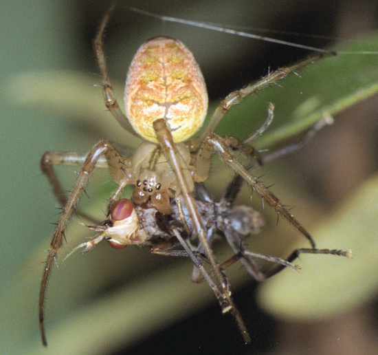
Meta segmentata consuming a fly
Feeding and digestion
The spider catches its prey with the appendages i.e. the legs. Then it
uses its sharp fangs to inject the venom into the prey. This venom also
contains enzymes that dissolve the prey.
The digestion is partly external in the prey itself. The liquefied
contents are sucked into the alimentary canal by the sucking stomach that is
located in the cephalothorax. The particles in the food are filtered
out by means of many hairs around and in the mouth. The filtering is so
efficient that it can separate the particles in Indian ink from the liquid.
From the sucking stomach the food is transferred via the intestine to the
midgut which is located in the abdomen where secretory cells
release enzymes which digest the food further.
Resorptive cells take up tiny globules of food and continue the
digestion. In this process, the food is broken down to a molecular size. The
midgut finally ends in the cloacal chambers where the faeces are
stored.
Breathing
The breathing system of spider is
different from ours. There are two separate systems involved, book lungs
and tracheae. The book lungs are assumed to be the first respiratory
system evolved which is later replaced by the tracheae.
A book lung consists of a stack of plates. Blood flows through these plates
and the gas is exchanged. The gases pass in and out by a diffusion process
and it therefore not very efficient. In more active spiders tracheae replace
part of the book lung or the whole book lung. The tracheae are tubes that do
not branch but run from the opening on the outside directly into the tissues
and organs.
Blood circulation
The spiders blood system is know as an open system. There is no blood
vessel system like in man but the blood pours out from the end of the
arteries directly onto the tissue where it runs freely. The cavity around
the heart is called pericardium.
The blood is different from man. The oxygen is bound to hemocyanin, a
molecule that contains copper, whereas in humans the oxygen is bound to
hemoglobin, a molecule that contains iron. The color of oxidized copper is
blue/green. Therefore, spiders have 'blue' blood. The blood contains some
cells, though to be mainly concerned with blood clothing, defense against
infection and wound healing. The blood pressure of spiders is high and is
with 0.18 atm nearly equal to men.
During the changing of the skin, this pressure is doubled. It is thought
that the blood pressure is used as a hydraulic force for walking.
Sensory organs
The spider processes different organs to communicate with the
environment. The most observable organs are the eyes and which were
described earlier.
Beside the eyes the spider has tactile sensors, tasting
sensors, slit organs and proprioreceptors. The tactile sensors are
the hairs that are spread all over the body. Movement of the hairs triggers
a nerve impulse. That this system works can easily be tested by shouting to
a spider. It will directly stop its activities because the sound waves move
the tactile hairs and the slit organs. Touching a spider will have the same
effect but that seems to be obvious.
The tasting sensors are located inside the mouth and on the end segments of
the palps. On the palps are hollow hairs into which volatile chemicals
enter. If a spiders manipulating its foods and touching it with its palps it
is tasting it!
The slit organs are spread around the outside of the body that senses
stress, vibration and gravitational forces. The proprioreceptors are found
on the appendages and are used to detect and to sense the position of the
legs and the direction of the movement.
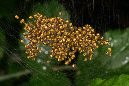
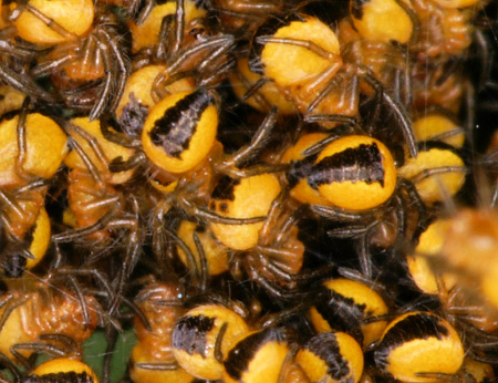
Spiderlings of Araneus species are clustered in a yellow ball that explodes on a slight disturbance.
Reproduction
The reproduction is a dangerous task for the male spider that he often has to pay for with his life. It is a complex process involving a wide range of approaches and tactics. The male has sperm production organs in it abdomen. Tubes lead to the genital opening that is located in the center of the epigastric furrow that can be found on the ventral side of the abdomen behind the legs. Before the male can mate, he has to transfer the sperms to the reservoirs in its palpal organs. To do this it makes a small web and a drop excluded from its genital opening. He then takes up the semen with its palp. The uptake of semen is probably a combination of capillary and gravitational forces. Then the dangerous part. He has to approach the female with the correct rituals. Failing these conditions is a death penalty. When the females accepts the male, the males connect its palp to the female epigyne and the semen is inserted.
The descendants
Once the eggs are fertilized and the eggs are matured, the female lays
her eggs. The mother has to take care of her unborn children because the
elements, predators or parasites can harm them. The Scytodes and the
Pholcus (daddy longleg) use the simplest form. They carry their children
in loose bundles with them. They can do that because they live indoors and
are not threatened by the weather. Other species like the wolf spiders put
their eggs in a sac of silk and attach the sac behind their body. After
their children are born, the female carries them for a while on her abdomen.
Many other species put their eggs in a sac of silk and guard them until
the youngsters emerge. Often she will care for her youngsters until she
dies. Among some species, the children eat the mother. Some Araneidae guard
their eggs until the mother dies in the autumn. The eggs stay unattended
until they hatch the following spring.
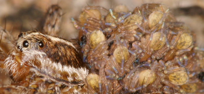
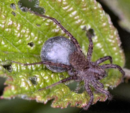
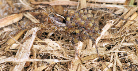
Wolf spider with cocoon attached to her spinners.
Wolf spider Pardosa with young spiders on her back
